
Scattered Brown Spots After Years of Arizona Sun
Sun spots are flat, UV-induced pigmented spots that surface where cumulative solar exposure has outpaced your skin's natural pigment-redistribution cycle. Clinically, the umbrella covers two related but distinct lesions: solar lentigines — the larger, sharply bordered tan-to-brown spots that build through adulthood — and ephelides (freckles) — the smaller, lighter spots that appear early in life and darken seasonally. Both are benign records of UV exposure, both sit in the upper dermis, and neither fades reliably on its own. In Scottsdale, where the UV index runs Very High to Extreme for most of the year, sun spots appear earlier and accumulate denser than in lower-UV climates.
The first decision is not which device — it is your Fitzpatrick skin tone. The correct path for lighter skin is fundamentally different from the safe path for medium-to-dark skin. At Desert Bloom, Dr. Natalya Borakowski, NMD assesses spot morphology, density, and Fitzpatrick type at consultation before any device or peel is selected. Sister concern: Age Spots — same clinical condition, used to describe the more isolated darker clusters that accumulate over decades; identical routing logic applies.
Lentigines, Ephelides, or PIH? Why the Distinction Matters
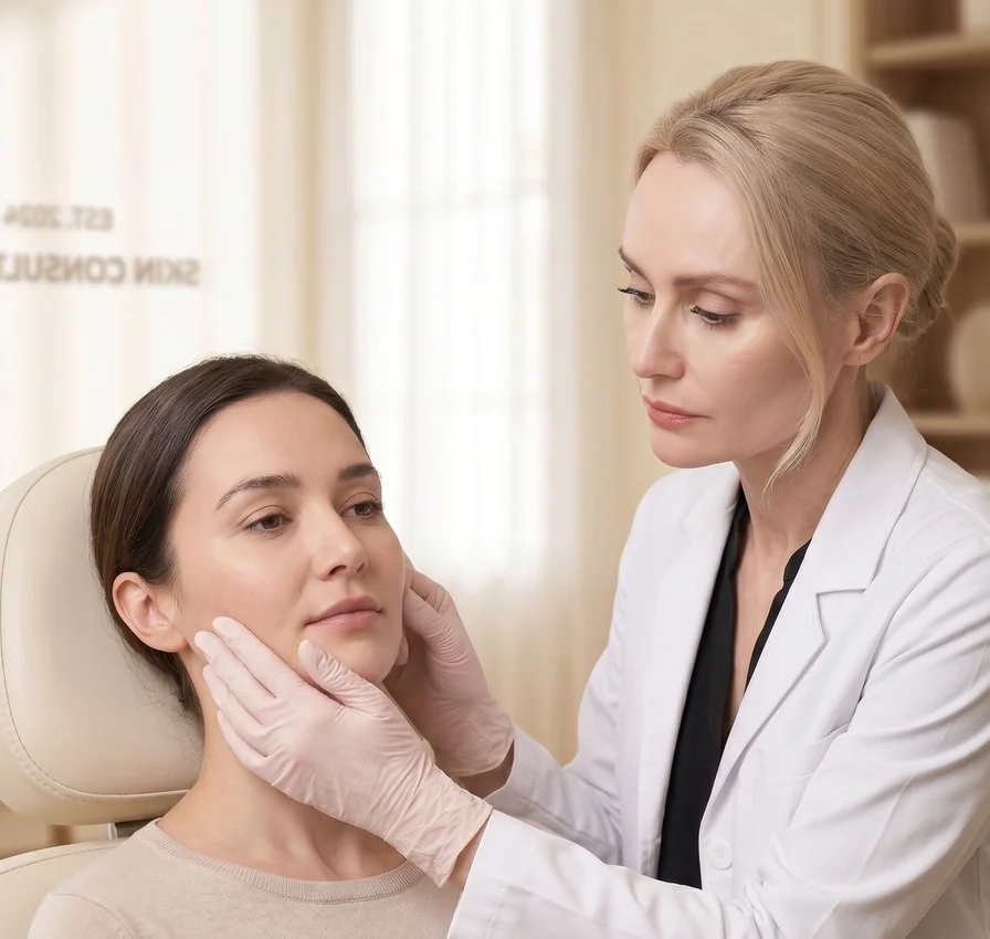
Most patients arrive having self-diagnosed. The problem is that solar lentigines (true sun spots — discrete, flat, sharply bordered, UV-driven, stable), ephelides (freckles — appear in childhood, fade seasonally, most prominent on Fitz I–II), and post-inflammatory hyperpigmentation (PIH — follows acne or injury, more persistent on medium-to-dark skin) all look like “brown spots” in a mirror but route to different treatments. Heat-based light applied to PIH, for example, can generate more PIH. Getting the type right before selecting a device is not a formality — it is the whole decision.
Sun Spot Treatments at Desert Bloom
Five in-clinic options across two Fitzpatrick-gated paths. Each card links to the full treatment page with procedure detail, candidacy, downtime, and Scottsdale pricing.

Fitz I–III · First-line
Photo Facial (Alexandrite 755nm)
Selective photothermolysis targets discrete melanin clusters from years of accumulated UV. Spots darken briefly over 7–14 days and shed. 1–3 sessions for most isolated spot patterns. Not for Fitz IV–VI.
All skin tones · First-line for Fitz IV–VI
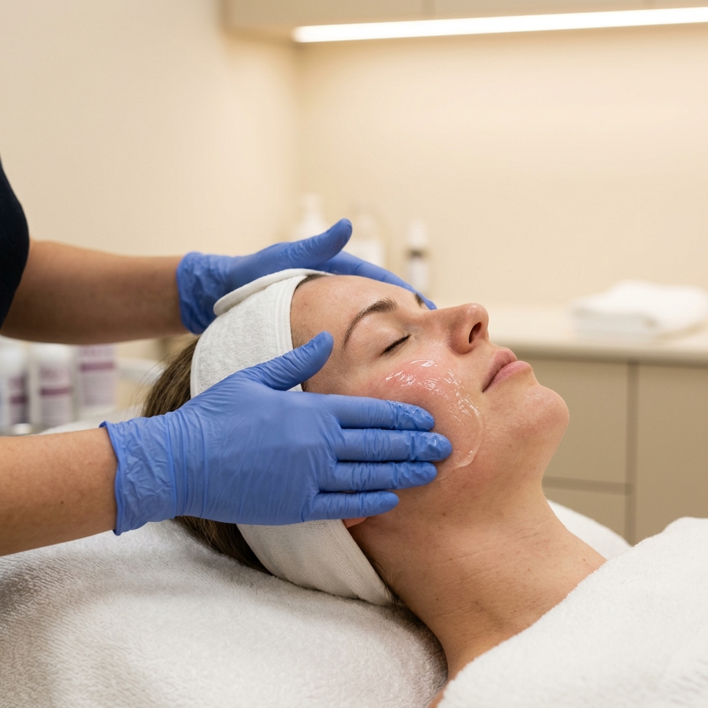
Unicorn Facial (PRX-T33)
Biorevitalization with TCA, hydrogen peroxide, and kojic acid. Suppresses excess melanin without ablation or thermal injury — no PIH risk. 3–5 days of light surface peeling; 3–5 sessions for full tone-evening.
All skin tones · Stepwise
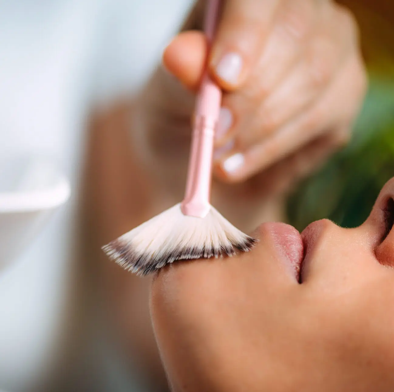
Custom Chemical Peel
Mandelic acid is selected for Fitz IV–VI (larger molecule, slower penetration, less PIH risk). TCA addresses moderate pigment on lighter skin. Used as primary correction, prep for Photo Facial, or maintenance. 3–7 days light peeling.
Fitz I–III · Deeper resurfacing
Erbium Laser Resurfacing
Ablative erbium for deeper photoaging where sun spots sit alongside texture and fine lines from accumulated UV damage. Single session with 5–10 days of focused recovery. Not appropriate for Fitz IV–VI pigment.
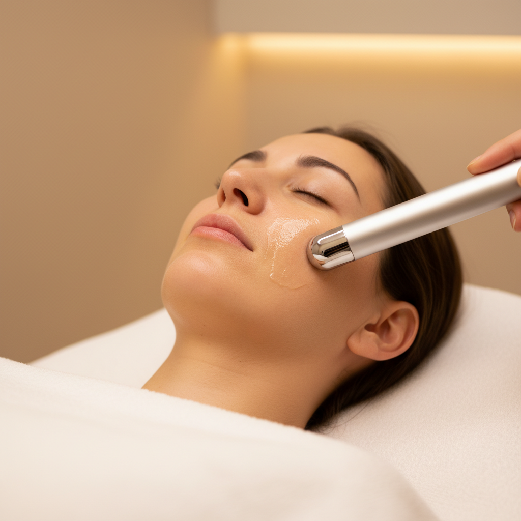
All skin tones · Zero downtime
Iontophoresis Facial
Low-current delivery of vitamin C, tranexamic acid, and niacinamide below the surface — the layer topicals alone cannot reach. Safe on all Fitzpatrick types. Best as monthly maintenance alongside primary correction; also suppresses new pigment formation.
Skin-tone gated routing
Which Path Is Safe for Your Skin?
Fitzpatrick skin type determines the safe device choice — not the size of the spots, not how long they've been there. Photo Facial is precise on lighter skin; on darker skin its melanin affinity becomes a liability, and the correct path is non-laser.
My skin is Fitzpatrick I–III (light, burns easily, sometimes tans)
→Start with Photo Facial — Alexandrite 755nm targets discrete melanin clusters with high precision. Chemical peels and iontophoresis support and maintain results between laser sessions.
Non-laser onlyMy skin is Fitzpatrick IV–VI (medium to dark, rarely burns)
→Start with Unicorn Facial — Non-laser path: PRX-T33 (Unicorn Facial) is first-line, supported by mandelic chemical peels and iontophoresis with brightening actives. No thermal risk, no PIH exposure.
When to See a Dermatologist First
True sun spots are flat, uniformly tan-to-brown, stable, and regularly bordered. Any pigmented lesion that fails the dermatology ABCDE rule needs dermatologist assessment to rule out lentigo maligna or other atypical pigmented lesions before any cosmetic treatment.
- A — Asymmetry: one half of the lesion differs from the other
- B — Border: ragged, notched, or irregular edges
- C — Color: multiple colors within a single lesion
- D — Diameter: larger than 6 mm
- E — Evolution: recent change in size, color, or texture
Bleeding, itching, or raised texture over a previously flat spot is also a reason to pause cosmetic treatment and seek a medical evaluation first.
At Desert Bloom, Dr. Borakowski reviews the morphology of every pigmented lesion at consultation and refers to a dermatologist when a spot shows any atypical features. Aesthetic treatment is not initiated on any lesion with a suspicious clinical history — that sequence keeps it safe.
Sun Spots — Frequently Asked Questions
What is the difference between sun spots and age spots?
What is the difference between sun spots and freckles?
How do sun spots differ from melasma?
Can sun spots be treated on darker skin tones?
Will sun spots come back after treatment?
How many sessions does sun spot treatment take?
How do I know if a dark spot needs a doctor before cosmetic treatment?

Medically reviewed by
Founder, Desert Bloom Skincare · 17 Years Experience
References
- 1.
Ortonne JP, Pandya AG, Lui H, Hexsel D. Treatment of solar lentigines. J Am Acad Dermatol; 2006;54(5 Suppl 2):S262-71.
- 2.
Passeron T, Picardo M. Melasma, a photoaging disorder. Pigment Cell Melanoma Res; 2018;31(4):461-465.
DOI: 10.1111/pcmr.12684
- 3.
Watanabe S. Basics of laser application to dermatology. Arch Dermatol Res; 2008;300 Suppl 1:S21-30.
- 4.
Grimes PE, Green BA, Wildnauer RH, Edison BL. The use of polyhydroxy acids (PHAs) in photoaged skin. Cutis; 2004;73(2 Suppl):3-13.
PMID 15002656
- 5.
Davis EC, Callender VD. Postinflammatory hyperpigmentation: a review of the epidemiology, clinical features, and treatment options in skin of color. J Clin Aesthet Dermatol; 2010;3(7):20-31.
PMID 20725554